Case 3 · IDH-mutant astrocytoma · Expert in the graph
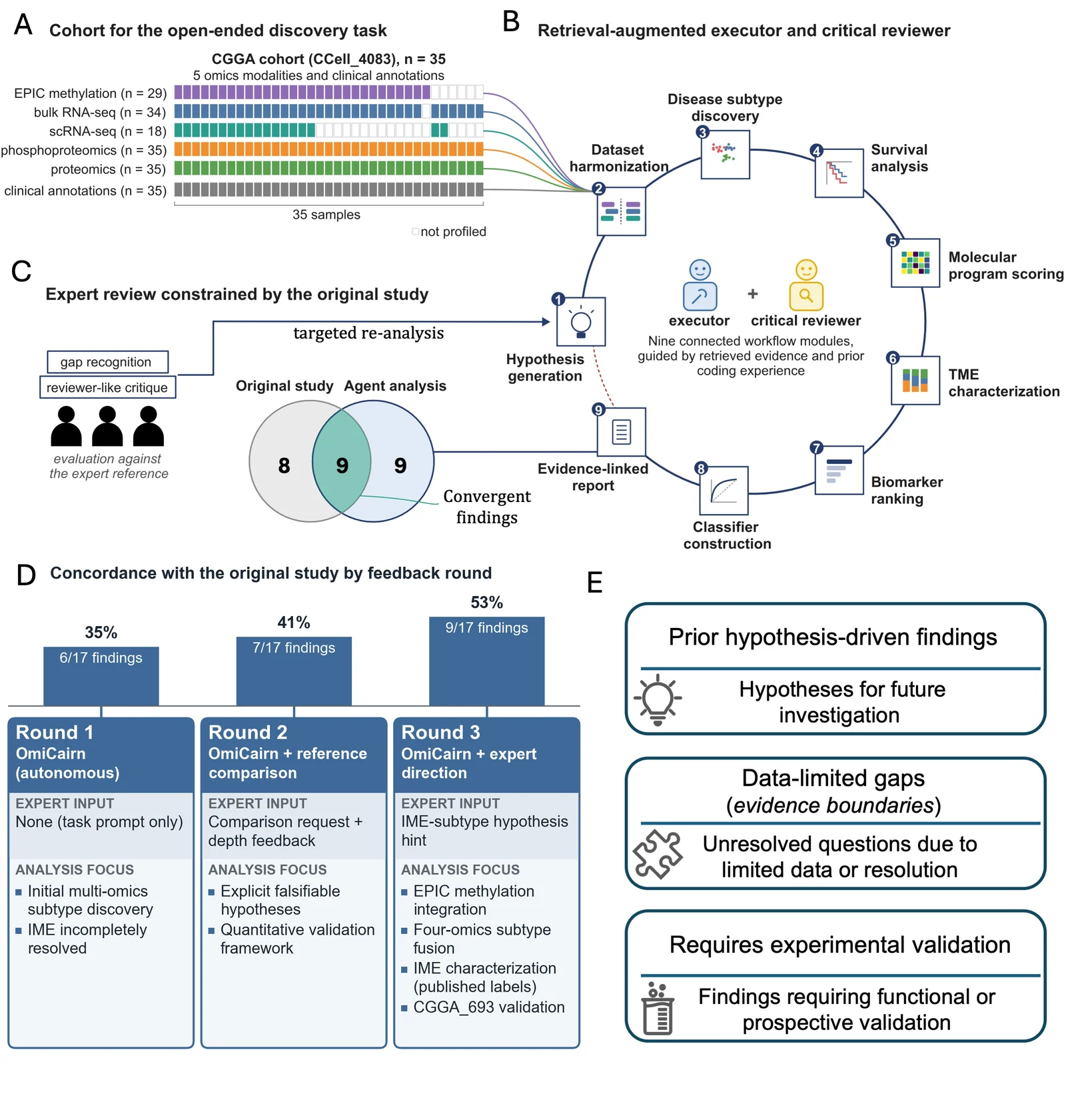
Three rounds of expert feedback raised concordance with a reference study from 6/17 to 9/17
OmiCairn analysed the publicly available CGGA subset (DataSet ID CCell_4083) of the IDH-mutant astrocytoma cohort reported by Tang et al. Each round was compared with 17 findings of the original study, with evaluators from the reference study team. The OmiCairn system stayed the same; only the human input grew.
Concordance by round
-
Round 1OmiCairn, autonomousExpert input: none (task prompt only)
6/17 35%
-
Round 2OmiCairn + reference comparisonExpert input: comparison request + feedback on its depth
7/17 41%
-
Round 3OmiCairn + expert directionExpert input: one hypothesis hint about the IME subtype
9/17 53%
reference finding recovered, including partial or trend-level recoveriesnot recovered
1 The data
35 tumours, five omics layers, uneven coverage
The dataset is the publicly available CGGA subset (DataSet ID CCell_4083) of the cohort reported by Tang et al.: 35 IDH-mutant astrocytoma samples with EPIC DNA methylation, bulk RNA-seq, single-cell RNA-seq, phosphoproteomics and proteomics, plus clinical annotations. Not every sample has every layer. Single-cell RNA-seq covers 18 of the 35.
No subtype structure or biomarker model was imposed before the analysis.
- Reference study
- Tang et al., Cancer Cell 2025, “Protein-based classification reveals an immune-hot subtype in IDH mutant astrocytoma with worse prognosis” doi
- Evaluators
- Members of the reference study team
- Comparison set
- A fixed set of 17 findings of the reference study
- External cohort
- CGGA_693 (RNA-level)
CGGA CCell_4083 · 35 samples · filled = profiled
2 The prompt
An open-ended question, with rules against assuming the answer
OmiCairn was asked to act as both an executor and a critical reviewer, and to explore the molecular and cellular mechanisms behind tumour heterogeneity, progression, recurrence and treatment resistance. The task covered subtype discovery, the tumour microenvironment, clinical associations and biomarker prioritization. It did not name the reference study’s subtypes or biomarkers.
The prompt also asked for spatial and longitudinal analyses where such data existed. The public subset has neither, which matters for what could and could not be recovered.
From the task prompt
“This project is data-driven. Do not assume any subtype, pathway, biomarker, or mechanism before analysis.”
“Treat associations as hypothesis-generating unless independently validated.”
“Report alternative explanations and negative findings.”
3 The workflow
Nine connected modules, run by an executor and a critical reviewer
OmiCairn organized the analysis itself. A reviewer step that recognises gaps against the reference sends the work back to hypothesis generation, which closes the loop between rounds.
- 1Hypothesis generation
- 2Dataset harmonization
- 3Disease subtype discovery
- 4Survival analysis
- 5Molecular program scoring
- 6Tumour microenvironment characterization
- 7Biomarker ranking
- 8Classifier construction
- 9Evidence-linked report
Expert review against the reference study → gap recognition → targeted re-analysis from module 1
7
candidate hypotheses tested
66
function blocks
40
implementation or data-handling issues resolved automatically
103
figures
123
tables
110 min
average runtime per round
Totals across the three rounds. Human input was the task specification, one comparison request with feedback on its depth, and one hypothesis hint; some manual debugging was still needed.
4 The three rounds
What the expert said, and what OmiCairn did with it
Each round reused the same OmiCairn system. What changed was the human input before the round started.
-
Round 1
OmiCairn, autonomous
6/17 35%
Expert input
None. Only the task prompt.
What OmiCairn did
- First multi-omics subtype discovery
- The immune-hot IME subtype was not resolved; it stayed merged with the AFM subtype
Recovered without any feedback
The adverse prognosis of the PPR subtype, and transfer of the subtypes across cohorts.
-
Round 2
OmiCairn + reference comparison
7/17 41% +1
Expert input
Compare your outputs with the original study. Then: the comparison lacks depth.
What OmiCairn did
- Rebuilt the workflow around explicit, falsifiable hypotheses
- Added a quantitative validation framework
Change
One more finding at partial level: a GBP2 trend toward the interferon program of IME.
-
Round 3
OmiCairn + expert direction
9/17 53% +2
Expert input
The question was resubmitted with a hint: a proteomics-defined IME subtype may contribute to recurrence.
What OmiCairn did
- Integrated the EPIC methylation data
- Fused four omics layers for subtyping
- Characterized IME using the published subtype labels
- Validated subtype signatures in the external CGGA_693 cohort
Change
Two more findings: the plasma-cell signal in IME (trend only) and the worse prognosis of IME (external cohort). Three others moved from partial or trend level to full recovery: the interferon program, T-cell exhaustion and cell-type composition.
5 Finding by finding
All 17 reference findings across the three rounds
Status per round from the case’s supplementary comparison. Counting every recovered, partial or trend-level cell gives exactly 6, 7 and 9, the totals in Figure 7D.
- Recovered
- Partial
- Trend only
- Discordant
- Not recovered
| Reference finding | Round 1 | Round 2 | Round 3 | Note |
|---|---|---|---|---|
| 1Four protein-based subtypes (AFM, NEU, PPR, IME) | Partial | Partial | Partial | Rounds 1–2 found three clusters with IME merged into AFM. By Round 3, AFM, NEU and PPR were supported; IME was only partly recovered by de novo clustering. (The supplementary tracking table counts Round 3 as confirmed; this page follows the more cautious manuscript text.) |
| 2PPR subtype has the worst prognosis | Recovered | Recovered | Recovered | Recovered without any feedback in Round 1. |
| 3A GBP1 / interferon program marks IME | Not recovered | Partial | Recovered | Round 2: GBP2 trend only. Round 3: tested with the published subtype labels. |
| 4T-cell exhaustion markers are enriched in IME | Trend | Trend | Recovered | Trend-level until Round 3, which used the published subtype labels. |
| 5Plasma cells / IGKC are highest in IME | Discordant | Discordant | Trend | Rounds 1–2 placed the signal in AFM (IME was merged into AFM). Round 3, with published labels: trend only, not significant with 4 IME samples. |
| 6IME carries a worse prognosis | Not recovered | Not recovered | Recovered | Not testable in Round 1 (IME merged); not significant in Round 2 (4 IME samples). Round 3: recovered in the external CGGA_693 cohort. |
| 7Cell-type composition patterns | Partial | Partial | Recovered | From bulk deconvolution; the single-cell data could not be used. |
| 8Pathway programs per subtype | Recovered | Recovered | Recovered | |
| 9Subtypes transfer across cohorts | Recovered | Recovered | Recovered | Recovered without any feedback in Round 1. |
| Not recovered in any round (8) | ||||
| 10Single-cell cell states (AC / OPC / MES / NPC) | Not recovered in any round | Data not available or not retrievable | ||
| 11CytoTRACE differentiation trajectory | Not recovered in any round | Data not available or not retrievable | ||
| 12Gemistocyte histology | Not recovered in any round | Data not available or not retrievable | ||
| 13Visium HD spatial organisation | Not recovered in any round | Data not available or not retrievable | ||
| 14Longitudinal evolution across matched primary and recurrent tumours | Not recovered in any round | Data not available or not retrievable | ||
| 15Protein-level validation in an independent proteomics cohort | Not recovered in any round | Data not available or not retrievable | ||
| 16MOFA latent factors | Not recovered in any round | Different method chosen | ||
| 17The reference study’s tree-based multi-modal classifier | Not recovered in any round | Different method chosen | ||
| Counted as concordant | 6/17 | 7/17 | 9/17 | |
6 Final overlap after Round 3
9 shared, 8 not recovered, 9 found only by OmiCairn
Shared (9)
- Four protein-based subtypes (AFM, NEU, PPR, IME)
- PPR subtype has the worst prognosis
- A GBP1 / interferon program marks IME
- T-cell exhaustion markers are enriched in IME
- Plasma cells / IGKC are highest in IME
- IME carries a worse prognosis
- Cell-type composition patterns
- Pathway programs per subtype
- Subtypes transfer across cohorts
Found only by OmiCairn (9)
- Kinase activity inferred from the phosphoproteomics
- A leave-one-omic-out stability analysis of the subtypes
- Modest RNA–protein concordance across genes
- Published RNA transcriptional subtypes did not stratify survival in this cohort (a negative result)
- A set of seven competing hypotheses with pre-stated support and refutation criteria
- A five-axis biomarker and target ranking
- Four-omic fusion with EPIC methylation (modest improvement)
- A bulk gemistocytic marker score associated with overall survival
- External validation of a four-subtype signature in CGGA_693
Not reported by the reference study and not independently validated. Treat as hypotheses.
What OmiCairn could not recover, and why
According to the manuscript, most of the eight unrecovered findings depended on data that were unavailable or could not be retrieved, or on different methodological choices. The per-finding reasons below come from the case’s supplementary comparison.
6 depended on data
- Single-cell cell states (AC / OPC / MES / NPC). Needs the single-cell data. Single-cell RNA-seq exists for 18 of the 35 samples, but the download failed in every round (incomplete or corrupted file).
- CytoTRACE differentiation trajectory. Same single-cell retrieval failure.
- Gemistocyte histology. Needs whole-slide images and spatial data, which were not used. A bulk marker score was computed instead (see below).
- Visium HD spatial organisation. Spatial transcriptomics is not part of the public CCell_4083 subset.
- Longitudinal evolution across matched primary and recurrent tumours. The public subset has one time point per patient.
- Protein-level validation in an independent proteomics cohort. No independent proteomics cohort was publicly available; OmiCairn validated at the RNA level in CGGA_693 instead.
2 reflect a different method
- MOFA latent factors. OmiCairn integrated the omics layers with a similarity-network approach instead.
- The reference study’s tree-based multi-modal classifier. OmiCairn built a signature-based classifier instead.
A different method is a choice, not a missing data type. Whether the reference study’s methods would give the same answer on this subset was not tested.
Where OmiCairn and the reference disagreed
In Rounds 1 and 2 the plasma-cell signal appeared highest in the AFM subtype rather than IME; the supplementary comparison attributes this to IME not yet being separated from AFM. With the published labels in Round 3 it moved to IME as a non-significant trend. Round 3 also produced a bulk gemistocytic marker score that was highest in PPR rather than IME. The reference study linked gemistocytes to IME using histology and single-cell data, which OmiCairn could not use, so the two observations rest on different evidence.
7 External cohort
Subtype signatures tested in CGGA_693
In Round 3, OmiCairn applied a four-subtype expression signature to the independent CGGA_693 cohort. Tumours assigned to PPR and to IME both had worse survival there.
This validation is at the RNA level. The reference study validated its protein-based classification in an independent proteomics cohort, which was not publicly available, so the two validations are not equivalent.
- Cohort
- CGGA_693, n = 237 with the signature applied
- IME
- HR 1.96 (95% CI 1.29–2.97), p = 0.0015
- PPR
- HR 2.38, p < 0.0001
- Four classes
- log-rank p < 0.0001
- Level
- Associative. No functional or prospective validation.
8 Evidence boundaries
OmiCairn sorted its own claims by strength of support
Findings aligned with the reference were kept apart from three weaker layers. All findings are associative, not causal.
Prior hypothesis-driven findings
Hypotheses for future investigation.
Data-limited gaps
Questions left unresolved by limited data or resolution.
Requires experimental validation
Findings that need functional or prospective validation.
9 Figure
Figure 7
10 Sources
Data and sources
- Dataset
- Publicly available CGGA subset, DataSet ID CCell_4083 (35 samples), of the cohort reported by Tang et al.
- External cohort
- CGGA_693
- Reference study
- Tang J. et al. Protein-based classification reveals an immune-hot subtype in IDH mutant astrocytoma with worse prognosis. Cancer Cell 43(11), 2136–2155.e14 (2025). doi:10.1016/j.ccell.2025.08.006
- This case
- The OmiCairn manuscript (in preparation): Results, Case 3, and Figure 7. Per-finding statuses, reasons for gaps, OmiCairn-only findings and external-cohort statistics come from the case’s supplementary materials.
- Status
- Hypothesis-generating. The evaluation was done with members of the reference study team.